
Description
Human iPSC‑derived retinal organoid models for a reproducible and reliable workflow
- Retinal organoids are three-dimensional structures derived from iPSCs that mimic the cellular composition and multi-layer architecture of the human retina
- Retinal organoids represent a physiologically relevant tool to study retinal development, disease mechanisms, toxicity, and potential therapeutic interventions in a highly controlled environment and with high translatability to human
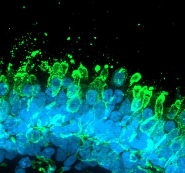
iPSC-derived retinal organoids contain the major retinal cell types organized in a laminar structure, as shown by the expression of Recoverin (photoreceptors), Rhodopsin (rods), Opsin LW/MW and SW (cones), PKC-α (bipolar cells), AP-2α (amacrine cells), SNCG (ganglion cells), CRALBP (Müller glial cells), and PROX1 (horizontal cells).

Kits
| Product name | Kit code | Maturation |
| Human iPSC-derived retinal organoids (unaffected, day 60), media, plate and pipettes kit | ax8006 | 60 days |
| Human iPSC-derived retinal organoids (unaffected, day 150), media, plate and pipettes kit | ax8007 | 150 days |
| Human iPSC-derived retinal organoids (unaffected, day 180), media, plate and pipettes kit | ax8010 | 180 days |
Media
| Product name | Product Code |
| Retinal organoid media | ax8009 |
Need a quote for this single product or for the whole cell + media + reagent kit? We’re here to help! Click the button below or send an email to operations@axolbio.com
Get a quote
Product highlights
- Derived from human iPSCs
- Contain all major retinal cell types, including rods, cones, bipolar, horizontal, amacrine, Müller glia, and ganglion cells
- Functional and respond to light as a result of the formation of photoreceptor outer segments
- Stratified, cell layers allow drug permeation
- Suitable for early toxicology workflows, developmental retina research, and drug discovery
Retinal organoids are fully characterised and monitored through the analysis of biomarkers specific for each cell type. For example, for cone photoreceptors, we monitor the expression of OPNSW, OPNMW, OPNLW, ARR3, RXRG; for rod photoreceptors we use RHO and NRL and for retinal ganglion cells we follow MATH5 (ATOH7) and BRN3 (POU4F2). Our organoids are scientifically validated to carry out advanced in vitro assays for many applications.
Depending on the cell type of interest, the organoids can be used at different stages of development
- Retinal ganglion cells are more abundant at day 60
- At day 150 the organoids contain major retinal cell types organized in a laminated manner
- Bipolar cells, cone and rod photoreceptors being more prevalent from day 180

Phenotypic characterization: expression of retinal organoid markers
iPSC-derived retinal organoids contain the major retinal cell types organized in a laminar structure, as shown by the expression of Recoverin (photoreceptors), Rhodopsin (rods), Opsin LW/MW and SW (cones), PKC-α (bipolar cells), AP-2α (amacrine cells), SNCG (ganglion cells), CRALBP (Müller glial cells), and PROX1 (horizontal cells).


Light response of the retinal organoids
Light-driven spiking activity recorded from presumed ON-Centre retinal ganglion cells (RGCs) and OFF-Centre RGCs . In the raster plot, each small vertical bar indicates the time stamp of a spike, where each row represents a different RGC . The left half illustrates the activity before stimulus onset and separated by the red line, the right half the activity when exposed to light .

Retinal organoids respond to compounds known to induce retinal toxicity in a dose-response manner
Assay overview

Drug treatment effect on retinal cell viability
Dose-response curve demonstrating the dose-dependent effect of known cytotoxic drug to the retina, on retinal organoids.
(A) Organoid health was monitored under the brightfield microscope which showed darker organoids with rougher edges following Thioridazine drug treatment. (B) Cell viability was measured as the percentage of ATP released in treated organoids relative to untreated organoids across increasing drug concentrations. The datapoints represent the average from two experimental repeats(n=2 independent experiments) conducted with 5 biological replicates per dose (n=5 organoids per dose). Day 230 organoids were treated for 24 or 72 hours with the indicated range of compounds concentrations.


Retinal organoids provide a versatile and human‑relevant platform for gene therapy vector assessment, enabling evaluation of AAV tropism, transduction efficiency, and promoter performance within photoreceptor‑like cells and other retinal lineages. Their complex, laminar organisation also supports disease modelling with advanced retinal organoid systems, accurately reflecting human retinal structure and developmental timelines for studying inherited and degenerative retinal disorders. In addition, these organoids are routinely used for investigational drug safety and efficacy, offering a responsive tissue model for cytotoxicity assays, functional readouts, and early preclinical screening relevant to retinal toxicology and therapeutic development.
Guided by over a decade of expertise, we ensure confidence in the quality of our iPSC-derived cells with rigorous QC. All cells come with a full Certificate of Analysis to ensure ethical and biological conformity for your peace of mind.
Axol has obtained all relevant licenses for commercial use.
Patient samples used to create these organoids have been ethically sourced and consented for research and commercial use. All cells come with a full Certificate of Analysis and are officially certified by HPSCreg®, to ensure ethical and biological conformity for your peace of mind.
- Li et al., 2025 (Cell Death & Disease) – Applied human iPSC‑derived retinal organoids to investigate molecular mechanisms underlying retinal degeneration, demonstrating the utility of 3D organoid systems for modelling disease‑relevant pathways.
https://www.nature.com/articles/s41419-025-07420-7 - Nakayama et al., 2024 (Journal of Cellular and Molecular Medicine) – Used retinal organoids to examine photoreceptor development and stress‑related responses, supporting their use as reproducible in vitro platforms for mechanistic and drug‑response studies.
https://onlinelibrary.wiley.com/doi/full/10.1111/jcmm.17670 - McClements et al., 2022 (Newcells Biotech) – Characterised AAV vector tropism in photoreceptor‑like cells within human iPSC‑derived retinal organoids, highlighting the relevance of organoid systems for evaluating gene‑therapy vector delivery and performance.
https://newcellsbiotech.co.uk/wp-content/uploads/2022/10/McClements_2022_AAV-tropism-in-retinal-organoids.pdf
- Retinal organoid media