
Receiving and maintaining sebocytes
General Notes
- Each lot is tested for absence of mycoplasma, HBV, HCV, HIV1/2.
- Expiration: guaranteed for up to 12 months from date of receipt if properly stored. Use cells immediately after thawing.
Storage
Sebocytes should be kept below -135°C, either in a deepfreezer (-145°C) or in the vapor phase of liquid nitrogen. Long-term storage at -80°C is not recommended. RPE cells are provided in CryoStor® CS10 cryopreservation medium (StemCell Technologies, e.g. #07959). CS10 contains 10% DMSO.
PhenoCULT®-SEB basal medium can be stored at 4°C for 1 month.
Supplements A and M should be stored at -20°C (or -80°C for long term) upon receipt. After thawing, supplements can be aliquoted and frozen, but should only be freeze/thawed once. Supplements can be maintained at room temperature for a maximum of 5 days.
Product use
Sebocytes are intended for in vitro research use only and are not to be used for any other purpose, which includes but is not limited to, unauthorized commercial uses, in vitro diagnostic uses, ex vivo or in vivo therapeutic uses or any type of consumption or application to humans or animals.
Safety precautions
Wear the appropriate personal protection equipment and handle the frozen vials with due caution. This product should be treated as potentially infectious and only used in adequate biological safety premises and conditions.
Do not ingest. In case of contact with eyes, rinse immediately with water for at least 15 min and seek medical advice.
Environmental measures: soak up with inert absorbent material. Clean with bleach and rinse thoroughly. Prevent further leakage or spillage if safe to do so.
Axol Bioscience cannot be held liable for any damage or losses resulting from the handling or from contact with the product.
Before you start
If you perform sebocyte culture for the first time, you might feel more confident with a little help. Our skilled technical support staff is fully available at support@axolbio.com and by phone or online at www.axolbio.com. Do not hesitate to contact us to get personalized help and fully achieve your goals with sebocytes. Axol Bioscience cannot guarantee the biological function or any other properties associated with performance of the product in researchers’ individual culture systems. Axol Bioscience guarantees that the product will meet the specifications only when assessed immediately after thawing using the recommended Protocol.
FOR RESEARCH USE ONLY
Not intended for human or animal diagnostic, therapeutic or clinical applications.
Procedure
Important notice: This protocol has been validated using the reagents and medium references mentioned. All steps should be performed in a sterile culture environment using appropriate handling procedures. Sebocytes are human cells and must be handled according to required ethical and safety rules.
- If your experiments require amplification of sebocytes with LOW lipid content, go to Method A on page 4.
- If your experiments require sebocytes with HIGH lipid content, go to Method M on page 6.
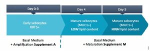
The chart below shows the timeline to obtain both stages of maturation:

Method A – Amplification of sebocytes with low lipid levels
Thawing
Reagents and medium
PhenoCULT®‑SEB sebocyte basal culture medium, containing:
- DMEM‑GlutaMAX™
- Ham’s F‑12
- Fetal Bovine Serum
- Human insulin
- Recombinant EGF
- Human hydrocortisone
- Adenine
- Cholera toxin B subunit
- Supplement A (“Amplification”, green cap). (in kit)
Store supplements at –20°C (or –80°C for long‑term) upon receipt.
After thawing, supplements can be aliquoted and frozen/thawed once.
Alternatively, they can be maintained at room temperature for a maximum of 5 days.
- Fibronectin (Sigma, cat. #F1141) (not included)
If you are performing this protocol for the first time, please make sure to use this specific reference.
Procedure
- Prepare tissue culture plates coated with fibronectin diluted to 1/100 in PBS.
Incubate for at least 2 h in a humidified incubator at 37°C (use 0.1 mL/cm² culture surface). - Pre‑warm a sufficient volume of PhenoCULT®‑SEB basal medium at 37°C
(2 mL for 10 cm² culture surface). - Thaw the cells:
- Transfer the vial of cells from storage by transporting the vial buried in dry ice.
- Remove the vial from dry ice and transfer it to a 37°C water bath.
- Do not submerge the vial.
- Remove the vial before the last bit of ice has melted (1–2 minutes).
- Do not vortex.
- Wipe out the outside of the vial with 70% ethanol and transfer it to a biological safety cabinet.
- Transfer the cells to 6 mL of PhenoCULT®‑SEB basal medium.
- Centrifuge at 290 g for 3 min, discard the supernatant, and resuspend in 2 mL of PhenoCULT®‑SEB basal medium supplemented with 1:1000 Supplement A.
- Perform a cell count to determine the number of viable cells.
- Remove the fibronectin solution from the culture plate and directly plate the sebocytes at 25,000 viable cells/cm².
Use 2 mL PhenoCULT®‑SEB medium + 1:1000 Supplement A per 10 cm² of culture surface. - Place the plate into a humidified incubator (37°C, 5% CO₂).
To ensure even plating, gently rock the culture plate back and forth and side‑to‑side twice. - Use PhenoCULT®‑SEB basal medium + 1:1000 Supplement A for at least 3 days after plating before use.
Keep the sebocytes in PhenoCULT®‑SEB with Supplement A for at least 3 days after plating before assays. - Cells can be passaged once when 90% confluence is reached (see page 7).
Method M – Maturation of sebocytes to reach high lipid levels
Thawing
Reagents and medium
PhenoCULT®‑SEB sebocyte basal culture medium, contains:
- DMEM‑GlutaMAX™
- Ham’s F‑12
- Fetal Bovine Serum
- Human insulin
- Recombinant EGF
- Human hydrocortisone
- Adenine
- Cholera toxin B subunit
- Supplement M (“Maturation”, yellow cap). Store supplements at −20°C (or −80°C for long term) upon receipt. After thawing, supplements can be aliquoted and frozen/thawed once. Alternatively, they can be maintained at room temperature for a maximum of 5 days.
- Fibronectin (Sigma, cat. #F1141) – If you are performing this protocol for the first time, please make sure to use this specific reference.
Procedure
- Prepare coated tissue culture plates with fibronectin diluted to 1/100 in 1x PBS. Incubate for at least 2 h in a humidified incubator at 37°C (use 0.1 mL/ cm2 cell culture surface).
- Pre‑warm a sufficient volume of PhenoCULT®‑SEB basal medium at 37°C (2 mL for 10 cm2 culture surface).
- Thaw the cells:
- Transfer the vial of cells from storage by transporting the vial buried in dry ice.
- Remove the vial from dry ice and transfer it to a 37°C water bath.
- Do not submerge the vial.
- Remove the vial before the last bit of ice has melted (1–2 minutes).
- Do not vortex.
- Wipe out the outside of the vial of cells with 70% ethanol and transfer to a biological safety cabinet.
- Transfer the cells to 6 mL of PhenoCULT®‑SEB basal medium.
- Centrifuge at 290g for 3 min, discard supernatant and resuspend in 2 mL of PhenoCULT®‑SEB basal medium supplemented with 1:1000 Supplement A.
- Perform a cell count to determine the number of viable cells.
- Remove the fibronectin solution from the culture plate and directly plate PCi‑SEB at a density of 25,000 viable cells/cm2. Use 2 mL PhenoCULT®‑SEB medium with 1:1000 Supplement A per 10 cm2 of culture surface.
- Place the plate into a humidified incubator (37°C, 5% CO2). To ensure an even plating, gently rock the culture plate back and forth and side‑to‑side twice.
- Use PhenoCULT®‑SEB basal medium supplemented with 1:1000 Supplement A for at least 3 days.
- Switch to PhenoCULT®‑SEB basal medium supplemented with 1:1000 Supplement M for the next 2 days until cells reach 90% confluence.
- Cells can be passaged once, but a notable amount of cell death might take place upon passaging as PCi‑SEB are lipid filled and fragile.
- Change medium every other day using 2 mL/10 cm2 culture surface (add 3 mL/10 cm2 culture surface for the weekend).
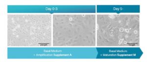
Note: One day after thawing, a significant amount of floating cells might be observed. This corresponds to normal cell death caused by our cell purification process. At D5 of culture, PCi‑SEB lipid droplets accumulate in the cytoplasm, while lipid‑filled cells detach from the culture plate and start holocrine secretion. As their physiological counterpart, fully mature PCi‑SEB will die after having released their content into the medium.
Expected morphology

Passaging
Human iPSC‑derived sebocytes can be passaged once after thawing. Passaging PCi‑SEB should be performed when the cells reach 90% confluency (usually 5–6 days after plating).
Reagents and medium
- PhenoCULT®‑SEB: Sebocyte basal medium (in kit)
- TrypLE™ Express (ThermoFischer, cat. #12605) (not included)
- Fibronectin (Sigma, cat. #F1141) – If you are performing this protocol for the first time, please make sure to use this specific reference. (not included)
Procedure
- Prepare tissue culture plates with fibronectin diluted 1/100 in 1x PBS (use 0.1 mL per cm² culture surface). Incubate for at least 2 h in a humidified incubator (37°C, 0.5% CO₂). Remove fibronectin solution before use.
- For PCi‑SEB maintenance, passage is usually performed when cells reach 90% confluence (within 5–6 days after plating). One can expect a yield of about 50,000 cells/cm². Do not overgrow PCi‑SEB, as it might impair survival after passage.
- Pre‑warm PhenoCULT®‑SEB medium and TrypLE™ Express.
- Discard culture medium from culture plates and briefly wash once with 1x PBS.
- Add 1 mL TrypLE™ Express per 10 cm² of culture surface.
- Incubate at 37°C for 5–10 min. Regularly check the cells, when all the cells look rounded, detach them by gently flushing the culture medium present in the plate.
- Transfer to a 15 mL sterile conical tube containing PhenoCULT®‑SEB medium (at least a 1/3 dilution ratio is necessary to stop TrypLE™ Express action).
- Gently centrifuge the cell suspension at 290 g for 3 min at room temperature. PCi‑SEB should form a visible pellet after centrifugation.
- Carefully remove the supernatant and re‑suspend the cell pellet in PhenoCULT®‑SEB medium with 1:1000 Supplement A.
- Perform a cell count to determine the number of viable cells and ensure optimal seeding density.
- Seed the cells on fibronectin‑coated culture surface at a density of 25,000 cells/cm² in PhenoCULT®‑SEB medium with 1:1000 Supplement A.
- Place the plate into the incubator.
To evenly distribute the cells, alternatively move the plate forward to backward and side‑to‑side twice. - Change medium every other day using 2 mL/10 cm² of culture surface (add 3 mL/10 cm² culture surface for week‑ends). Use Supplement M to push maturation, if needed.
- Cells can be assayed as early as 24 h after passage.