Modeling ALS with iPSC-derived motor neurons, microglia and astrocytes

Modeling ALS with iPSC-derived motor neurons, microglia and astrocytes

Authors: Helena Raine, Jessica Tilman, David Wallbank, Sian Humphreys, and Sapna Vyas

Introduction

Amyotrophic Lateral Sclerosis (ALS), or Lou Gehrig’s disease, is a neurodegenerative disease characterised by the progressive degeneration of motor neurons in the spinal cord and the motor cortex. This degeneration of neurons ultimately results in respiratory failure and death around 3 years after diagnosis¹. There is no current cure for ALS or effective treatments available.

ALS is linked to various mutations, with the C9orf72 expansion, TDP-43, and SOD1 most frequently implicated in familial ALS, although these only correspond to 5-10% of ALS cases. Sporadic ALS makes up the remaining 90 to 95% of all cases of the disease².

Slow progress in the field has in part been due to the heterogeneity within this disease, and due to the lack of appropriate in vitro models and platforms for drug discovery to gain insight into the pathophysiology of ALS. Research into ALS has been primarily focused on animal models, cell lines and patient material, and except for SOD1G37R mice3, animal models have failed to show translatability to clinical outputs. Patient samples and post-mortem material are not scalable or reproducible enough to provide a reliable platform for drug discovery, further complicated by the quick progression of the disease.

Human induced pluripotent stem cells (iPSCs) have emerged as a valuable tool for disease modeling and drug discovery in neurodegeneration, providing scalable, reproducible and human-relevant models. The ability to genetically manipulate cells to introduce and correct mutations as well as utilising patient derived cells further adds value to these models in the study of heterogeneous conditions such as ALS and can provide a method to study large cohorts of samples for patient stratification.

axoCells™ iPSC-derived motor neurons from donors affected by ALS backgrounds – C9orf72 expansion, TDP-43 A382T, SOD1 D109Y and a donor unaffected by the disease were used to develop a model to study the morphological and functional differences between the neurons.

Additionally, a more physiologically relevant tri-culture model of motor neurons, astrocytes, and microglia was developed to further interrogate the role of neuroinflammation and cell to cell signalling in neuronal dysfunction. Future work will focus on expanding this work to include sporadic ALS lines.

Methods and assays

axoLines™ iPSCs

Fibroblasts from unaffected and ALS donors were reprogrammed to iPSCs using Sendai Virus.

axoCells™ iPSC-derived cells

iPSCs were differentiated to motor neuron progenitors using small molecule induction methods prior to cryopreservation. axoCells™ iPSC-derived motor neurons were differentiated from donors affected by ALS backgrounds (C9Orf72 expansion (ax0074), TDP-43 A382T (ax0079), SOD1 D109Y (ax0735)) and an unaffected donor (ax0076). axoCells™ motor neuron progenitors were thawed and matured to motor neurons over 21 days and quality control performed by immunocytochemistry for expression of HB9, ChAT, Islet-1, SMI-32, and TUJ1, as well as functional firing on an Incucyte® Live-Cell Analysis System.

iPSC-derived microglia were also differentiated using a small molecule induction method and cryopreserved as immature microglia. axoCells™ microglia were thawed and matured over 7 days prior to assessing expression of IBA-1, TMEM119, and CX3CR1 by immunocytochemistry.

iPSC-derived astrocytes were differentiated using small molecule induction methods prior to cryopreserving. Once thawed, axoCells™ astrocytes were cultured for 2 days and assessed for expression of EAAT2, GFAP, Nestin, S100, ALDH1, AQP4 and TUJ1.

A tri-culture model including each cell type was also developed, which is now available as a complete kit (ax0194).

Immunocytochemistry

The cells were fixed using 4% paraformaldehyde at room temperature (RT) for 20 minutes and washed in phosphate buffered saline (PBS). Cells were then permeabilised using 0.1% Triton X and blocked in 5% Donkey Serum. Primary Antibody SMI-32 was used to stain the motor neurons overnight. Secondary antibodies conjugated to FITC were then added for a 2-hour incubation at RT, prior to imaging using a Leica microscope.

Incucyte® Neuronal Activity Assay

iPSC-derived motor neuron progenitor cells from unaffected and disease lines were thawed and seeded at 40,000 cells per well of a 96-well plate. The neurons were matured for 21 days. On Day 5 – 7, cells were transduced using the Incucyte® Neuroburst Orange Lentivirus Reagent (Neuroburst Orange; Sartorius, 4736) a live-cell neuronal labeling reagent driven by a synapsin promoter, resulting in the long-term expression of an orange fluorescent calcium indicator. Spontaneous activity monitored throughout maturation up to Day 21 using an Incucyte® Live-Cell Analysis System and integrated Incucyte® Neuronal Activity Analysis Software Module.

Quantification of TDP-43 aggregation

iPSC-derived motor neurons from unaffected and ALS (C9orf72 expansion) donors were thawed and cultured for 3 days prior to adding sodium (Na) arsenite as a stressor to induce TDP-43 mislocalisation/aggregation for 2 hours. Cells were then fixed as described above and stained for total TDP-43 and TUJ1. Cells were imaged and quantification of nuclear and cytoplasmic staining performed by Nikon.
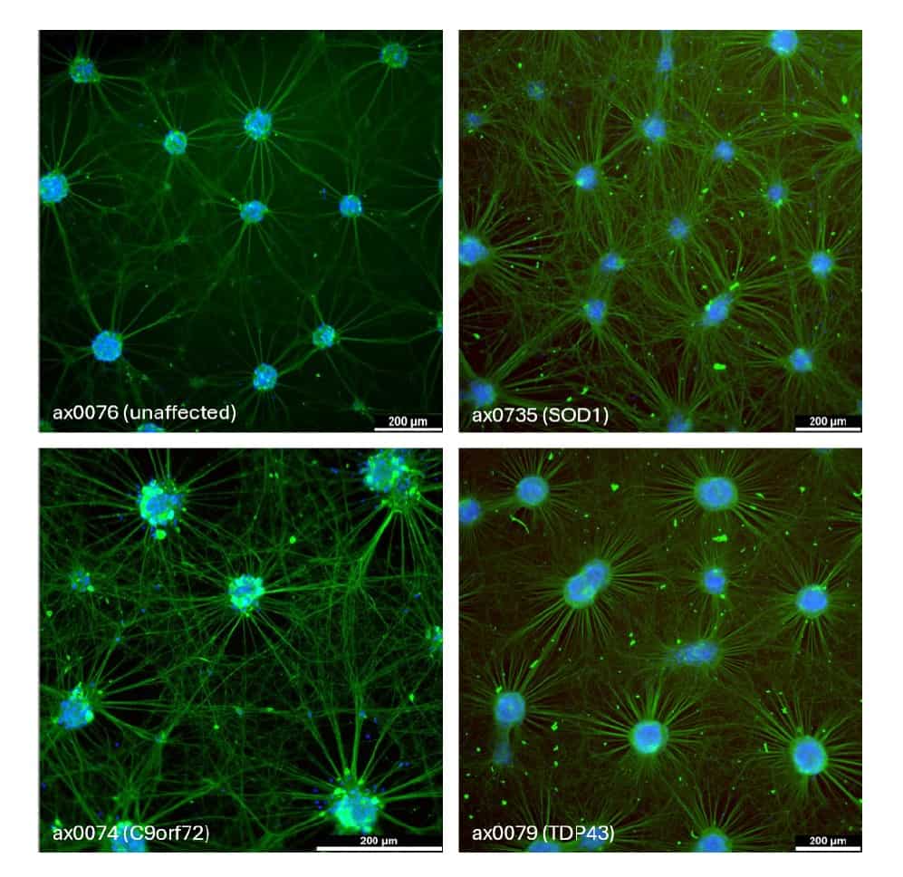
iPSC-derived motor neuron morphology

Figure 1. Immunocytochemistry staining of mature axoCells™ iPSC-derived motor neurons from an unaffected donor (ax0076) and ALS donors, C9orf72 expansion >145 G4C2 (ax0074), TDP-43 heterozygous A382T mutant (ax0079), SOD1 heterozygous D109Y mutant (ax0735). Cells were stained using a SMI-32 marker on day 21 of maturation.

Mature motor neurons demonstrate differences in morphology between the unaffected control cells and the various ALS derived motor neurons. During the 21-day maturation from progenitors, motor neurons form large clusters with thick neuronal processes connecting different bundles as well as smaller neurites present at the junctions.

Neurons from unaffected individual shows a uniform distribution of compact bundles. ALS motor neurons show a more disorganised neurite network, with different size bundles and higher proportion of thinner neurites. Differences in morphology and network disorganisation may in part drive decreased synchronicity and hyperexcitability in the ALS neurons.

iPSC-derived motor neuron activity

Figure 2. A) Representative image of fluorescent signalling displayed by healthy neurons monitored using the Incucyte® Neuronal Activity Assay showing network burst events and B) image showing calcium traces across the neuronal network.

Figure 3. Examples of calcium oscillation traces measured using the Incucyte® Neuronal Activity Assay recorded over 60 seconds of A) TDP-43 mutant iPSC-derived motor neurons and B) C9orf72 expansion iPSC-derived motor neurons.

Spontaneous neuronal activity was characterised using the Incucyte® Neuronal Activity Assay by transducing the cells with a flourescent calcium sensitive reporter, Incucyte® Neuroburst Orange, which is driven off the synapsin promoter. Activity can be measured daily to monitor changes in neuronal firing or patterns over the maturation period. By Day 21, motor neurons from unaffected individuals show highly synchronised and regular network bursting (Figure 2).

Motor neurons derived from ALS patients each show distinct firing patterns compared to control line, however a common feature is reduced synchronicity (mean correlation), a decreased number of active objects, reduced network burst rate, and reduced burst strength (Figures 3 – 4). Firing patterns have been replicated using a multi-electrode array (MEA) system to validate the disease phenotypes (data not shown).

Figure 4. A) Mean Burst Strength of motor neurons from an unaffected donor compared to 2 batches of motor neurons from donors with the TDP-43 mutation and the C9orf72 expansion mutation, respectively, over a 21-day maturation period measured using the Incucyte® Live-Cell Analysis System. Quantification of spontaneous neuronal activity on day 21 neurons from different patient backgrounds B) Burst Strength C) Burst Rate D)Mean Data represent 5-7 independent experiments. A paired One-Way ANOVA was performed to compare differences between the lines. *p<0.05, **p<0.01

Quantification of TDP-43 aggregation

TAR DNA-binding protein (TDP-43) pathology is of particular interest in ALS research as it is a shared feature across both familial and sporadic ALS, and more recently has been found to be present in other neurodegenerative conditions. TDP-43 is an RNA/ DNA binding protein involved in RNA transcription, splicing, stability, transport and translation, and is predominantly present in the nucleus. In ALS, TDP-43 becomes mislocalised to the cytoplasm where it is misfolded, ubiquitinated, and phosphorylated, resulting in protein aggregation.

Loss of nuclear TDP-43 leads to loss of function events, such as mis-splicing of cryptic exons, and presence in the cytoplasm drives toxicity4. Using iPSC-derived motor neurons to replicate this pathology has been challenging due to the difficulty in observing protein aggregation in models which do not fully recapitulate an ‘aged’ brain. In addition, the propensity for the neurons to form cell bundles makes identifying individual cells and nuclei challenging. To overcome this issue, cells were stained and imaged on Day 3 post thaw, and a stressor added (sodium (NA) arsenite) to induce mislocalisation (Figure 5).

Figure 5. A) Motor neuron progenitors from C9orf72 expansion line (ax0074) stained with TUJ1 (Green) and TDP-43 (Red). B) Quantification of TDP-43 in ALS or stressed control motor neurons in monoculture (n=1) per-formed by Nikon labs. C) The cytoplasmic to nuclear ratio of the TDP-43 in those neurons.

Development of tri-culture models

While studying motor neurons in monoculture provides valuable information on difference between control and affected neurons and can provide key information on effect of compound or new therapeutics on these cells, it does not address the role of cell-to-cell interactions. The role of both microglia and astrocytes in neurodegeneration and inflammation has been incresingly investigated.

To develop a more complex model, Axol developed a co-culture model with motor neurons, microglia and astrocytes, all derived from the same starting control iPSC line (ax0076, ax0664 and ax0704).

Multiple factors were tested, including cell densities, timepoints of addition and media composition with main endpoints of immunocytochemistry and spontaneous neuronal activity (Figure 6). Neuronal activity was assessed using the Incucyte® Neuronal Activity Assay to determine the effect of adding glial cells at different timepoints and different cell densities.

The kinetic data was used to confirm neuronal function and showed adding glial cells at an earlier timepoint had a larger effect on influencing neuronal firing compared to cell densities (Figure 6).

Figure 6. Immunocytochemistry staining of A) Motor neurons in monoculture stained with TUJ1 (white) B) Mo-tor neurons stained with TUJ1 (White), microglia stained with IBA-1 (Red) and CD44 (Green), astrocytes stained with CD44 (Green). Neuronal activity measured using the Incucyte® Neuronal Activity Assay over the matura-tion of motor neurons, astrocytes, and microglia highlighting differences in burst strength when glia are added at the early timepoint (~day 5) (C), the later timepoint (~day 12) (D) and at different densities.

Summary

In this study, motor neurons derived from iPSCs reprogrammed from donors diagnosed with ALS were shown to display morphological and physiological disease phenotypes. Using these cells to model ALS in vitro provides valuable insight into disease progression and treatments with key assay parameters including temporal assessment of neuronal activity and endpoint measurements of neurite outgrowth and TDP-43 aggregation.

References

  1. Gregory, M. et al. (2020) ‘Genetics of Amyotrophic Lateral Sclerosis’, Current Genetic Medicine Reports, 8, pp. 121–131. Doi: https://doi. org/10.1007/s40142-020-00194-8.
  2. Matteo Calafatti, Cocozza, , Limatola, C. and Garofalo, S. (2023). Microglial crosstalk with astrocytes and immune cells in amyotrophic lateral sclerosis. Frontiers in Immunology, 14. doi: https://doi.org/10.3389/fimmu.2023.1223096.
  3. Leenor Alfahel, Aleksandar Rajkovic, Adrian Israelson (2024) Protocol for handling and using SOD1 mice for amyotrophic lateral sclerosis pre-clinical studies, STAR Protocols, Volume 5, Issue 4, 103459, ISSN 2666-1667 doi: https://doi.org/10.1016/j.xpro.2024.103459.
  4. Genin, C. and Paquis-Flucklinger, V. (2026). ALS-related proteinopathies: From TDP-43 to mitochondrial proteinopathies. Current opinion in neurobiology, 97, p.103163. doi: https://doi.org/10.1016/j.conb.2025.103163.